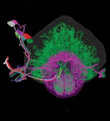
 In this video from SC19, Berkeley researchers visualizes an entire brain at nanoscale resolution. The work was published in the journal, Science.
At the core of the work is the combination of expansion microscopy and lattice light-sheet microscopy (ExLLSM) to capture large super-resolution image volumes of neural circuits using high-speed, nano-scale molecular microscopy.
In the simulation, 1.3 terabytes of multi-channel ExLLSM data show the 3D spatial relationships between essential proteins responsible for many functions in the brain including neural connectivity and signal regulation.
With its high speed, nanometric resolution, and ability to leverage genetically targeted, cell type-specific, and protein-specific fluorescence labeling, ExLLSM fills a valuable niche between the high throughput of conventional optical pipelines of neural anatomy and the ultrahigh resolution of corresponding EM pipelines,” the researchers stated in their paper.
Visualizing such large microscopy data has been a computational challenge for researchers, who are forced to create static view videos rather than being able to navigate through data. This impedes scientific discovery and takes many hours to generate preprocessed visuals.
To visualize the data, the team used NVIDIA’s IndeX platform, a 3D volumetric visualization SDK that allows scientists and researchers to visualize and interact with massive data sets in real-time regardless of the data size.
NVIDIA IndeX uses NVIDIA GPU clusters for providing instantaneous visual feedback, insights, and certainty. In this case, 6 NVIDIA DGX-1 systems, comprised of multiple NVIDIA V100 GPUs.
The technology should enable statistically rich, large-scale studies of neural development, sexual dimorphism, degree of stereotypy, and structural correlations to behavior or neural activity, all with molecular contrast,” the researchers stated.
Source: NVIDIA