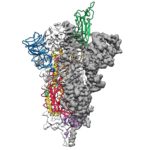
Researchers from The University of Texas at Austin and the National Institutes of Health have made a critical breakthrough toward developing a vaccine for the 2019 novel coronavirus by creating the first 3D atomic scale map of the part of the virus that attaches to and infects human cells. “Mapping this part, called the spike protein, is an essential step so researchers around the world can develop vaccines and antiviral drugs to combat the virus. The paper was published Feb. 19 in the journal Science.”
XSEDE Supercomputers Advance Skin Cancer Research
In this TACC podcast, UC Berkeley scientists describe how they are using powerful supercomputers to uncover the mechanism that activates cell mutations found in about 50 percent of melanomas. “The study’s computational challenges involved molecular dynamics simulations that modeled the protein at the atomic level, determining the forces of every atom on every other atom for a system of about 200,000 atoms at time steps of two femtoseconds.”
Video: How to Scale from Workstation through Cloud to HPC in Cryo-EM Processing
Lance Wilson from Monash University gave this talk at the GPU Technology Conference. “We’ll review the last two years of development in single-particle cryo-electron microscopy processing, with a focus on accelerated software, and discuss benchmarks and best practices for common software packages in this domain. Our talk will include videos and images of atomic resolution molecules and viruses that demonstrate our success in high-resolution imaging.”
Accelerating Cryo-EM with Intel Technologies
In this video from the Intel HPC Developer Conference, Erik Lindahl from Stockholm University describes the challenges of cryo-EM, a technique that fires beams of electrons at proteins that have been frozen in solution, to deduce the biomolecules’ structure. “Structural biology is going through a revolution where cryo-EM now determine 3D structures from 100,000s of noisy images, but it relies on very large computations. I will present our work with Intel to accelerate the RELION program with x86 SIMD, TBB, and MKL to provide outstanding performance.”
Bright Computing Powers SingleParticle.com for cryo-EM
Today Bright Computing announced a reseller agreement with San Diego-based SingleParticle.com. The company specializes in turn-key HPC infrastructure designed for high performance and low total cost of ownership (TCO), serving the global research community of cryo-electron microscopy (cryoEM). “With Bright, the management of an HPC cluster becomes very straightforward, empowering end users to administer their workloads, rather than relying on HPC experts,” said Dr. Clara Cai, Manager at SingleParticle.com. “We are confident that with Bright’s technology, our customers can maintain our turn-key cryoEM cluster with little to no prior HPC experience.”
Podcast: Mapping DNA at Near-Atomic Resolution with Cryo-EM
In this podcast, Berkeley Lab’s Eva Nogales describes how her team is using a new imaging technology that is yielding remarkably detailed 3-D models of complex biomolecules critical to DNA function. Using cryo-electron microscopy (cryo-EM), Nogales and her colleagues have resolved the structure at near-atomic resolutions of a human transcription factor used in gene expression and DNA repair.
Cryo-EM Moves Forward with $9.3M NIH Award
The National Institutes of Health (NIH) has awarded $9.3 million to the Department of Energy’s Lawrence Berkeley National Laboratory (Berkeley Lab) to support ongoing development of PHENIX, a software suite for solving three-dimensional macromolecular structures. “The impetus behind PHENIX is a desire to make the computational aspects of crystallography more automated, reducing human error and speeding solutions,” said PHENIX principal investigator Paul Adams, director of Berkeley Lab’s Molecular Biophysics and Integrated Bioimaging Division.”
Berkeley Lab Algorithm Boosts Resolution on Cryo–EM
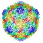
Berkeley Lab researchers have developed the first 3-D atomic-scale model of P22 virus that identifies the protein interactions crucial for its stability. “This is a great example of how to exploit electron microscopy technology and combine it with new computational methods to determine a bacteriophage’s structure,” said Paul Adams, Berkeley Lab’s Molecular Biophysics & Integrated Bioimaging division director and a co-author of the paper. “We developed the algorithms—the computational code—to optimize the atomic model so that it best fit the experimental data.”
Dell & Intel Collaborate on CryoEM on Intel Xeon Phi
In this video from SC16, Janet Morss from Dell EMC and Hugo Saleh from Intel discuss how the two companies collaborated on accelerating CryoEM. “Cryo-EM allows molecular samples to be studied in near-native states and down to nearly atomic resolutions. Studying the 3D structure of these biological specimens can lead to new insights into their functioning and interactions, especially with proteins and nucleic acids, and allows structural biologists to examine how alterations in their structures affect their functions. This information can be used in system biology research to understand the cell signaling network which is part of a complex communication system.”


